Drawings below are first made from photographs that are then compared with the preserved squid under a microscope. A small error will occur in identifying the two classes of photophores (red - counterilluminating photophores with red color filters = "complex photophores, and blue - simple photophores plus lens bearing photophores = "non-complex photophores." Blue photophores cannot, yet, be reliably separated, in preserved squid, into the two component types.). A damaged or developing/transforming photophore cannot always be reliably identified. Even the presence or absence of photophores can be difficult on strongly curved surfaces, for example, the sides of the head or mantle when viewed ventrally. Therefore, the photophore arrangement, sen below, is most accurate on the ventral surfaces when viewed ventrally and on the lateral surfaces when viewed laterally.

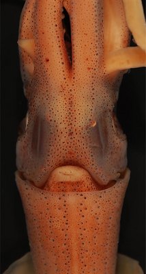
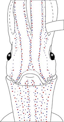
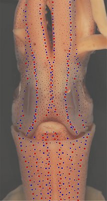
Figure. Ventral views of the integumental photophores of Abraliopsis sp. P, mature male, 26 mm ML, tropical South Pacific waters. Left - Photograph of the preserved squid. Middle - Outline drawing from photograph with all integumental photophores represented by colored dots. Right - Photograph with colored dots superimposed on integumental photophores. Red dots - Complex photophores. Blue dots - Non-complex photophores. Images by R. Young.
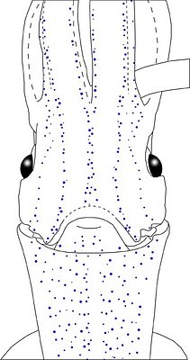
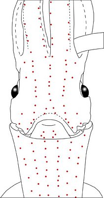
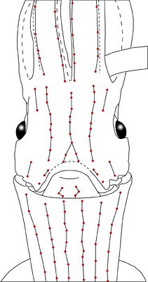
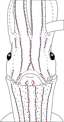
Figure. Same squid (26 mm ML), same view as above. Top left - Drawing showing only the non-complex (blue) photophores. Top middle - Drawing showing only the complex (red) photophores. Top right - Same drawing with lines connecting red photophores to aid comparisons of photophore patterns between species. Bottom - Drawing showing all photophores and lines showing red photophore patterns. Images by R. Young.
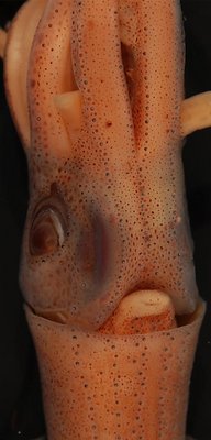
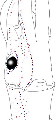
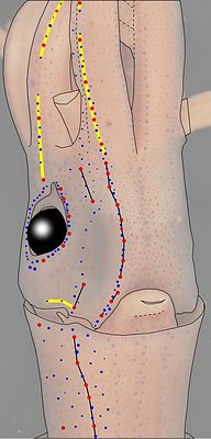
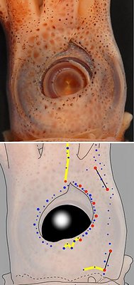
Figure. Views of the same squid showing lateral photophores. Left - Photograph, oblique view, of the preserved squid. Left middle - Drawing taken from the photograph showing distribution of lateral "red" and "blue" photophores. Right middle - Same drawing as previous one. Lines connecting photophores assist in comparing the organization of the photophores between species. Top right - Photograph, side view, of the preserved squid. Bottom right - Outline drawing over dimmed photograph with all integumental photophores represented by colored dots, and with lines connecting photophores. Red dots - Complex photophores. Blue dots - Non-complex photophores. Black lines - Patterns based on red photophores. Yellow lines - Patterns based on red and blue photophores. Images by R. Young.
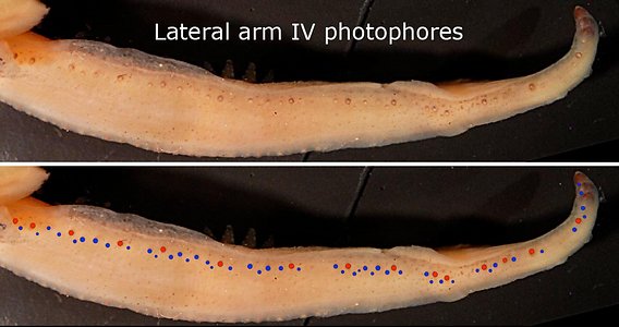
Figure. Dorso-aboral view of the lateral arm IV photophores of Abraliopsis sp. P, 28 mm ML, mature male. Top - Photograph of the preserved right arm IV; specimen old and chromatophores faded. Bottom - Same photograph with colored dots superimposed on photophores. Red dots - Complex photophores. Note that the photophores are discontinuous. Blue dots - Non-complex photophores. Images by R. Young.
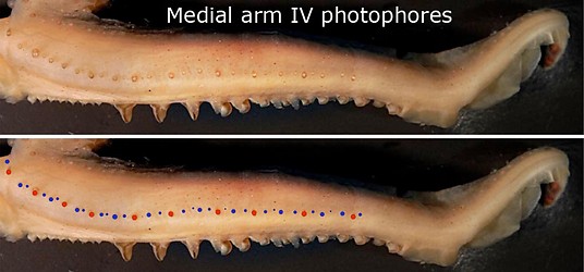
Figure. Ventro-aboral view of the medial arm photophores on the same arm as above. Top - Photograph of the preserved right arm IV; specimen old and chromatophores faded. Bottom - Same photograph with colored dots superimposed on photophores. Note that the photophores are continuous and that the series ends short of the proximal end of the ventral flap of the hecotcotylus. Red dots - Complex photophores. Blue dots - Non-complex photophores. Images by R. Young.
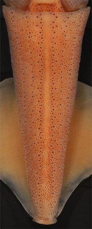
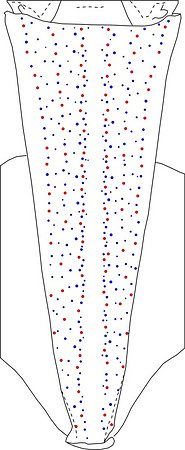
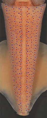
Figure. Mantle photophores of Abraliopsis sp. P, mature male, 26 mm ML, central Equatorial Pacific. Left - Photograph of the preserved squid. Middle - Outline drawing from the photograph with all integumental photophores represented by colored dots. Right - Photograph with dots superimposed dots on integumental photophores. Red dots - Complex photophores. Blue dots - Non-complex photophores. Images by R. Young.
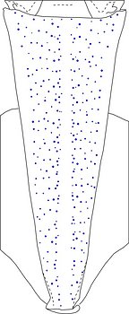
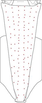
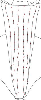
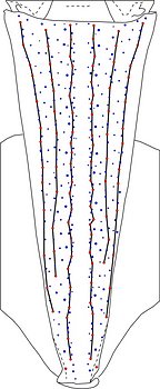
Figure. Ventral view of outline drawings of the same squid showing different photophore components. Left - Drawing showing only the non-complex (blue) photophores. Left middle - Drawing showing only the complex (red) photophores. Right middle - Same drawing with lines connecting red photophores to aid comparisons of photophore patterns between species. Right - Drawing showing all photophores and lines showing red photophore patterns. Images by R. Young.
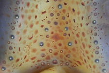
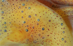
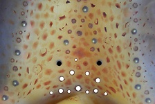
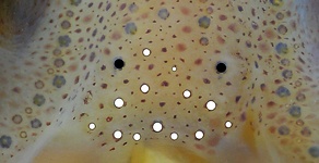
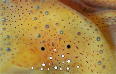
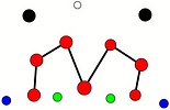
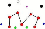
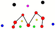
Table. Ventral view of the funnel-groove region of Abraliopsis sp. P. Top tier - Funnel groove showing photophores of funnel groove and surrounding tissue. Middle tier - In the photograph, white dots placed over the "white" photophores of the funnel groove. Black dots placed over the two most posteromedial "red" photophores of the ventral head to mark the anterolateral edges of the funnel groove. Bottom tier - Same pattern of dots as in photographs but dots are colored. Colored dots, lines - Aid in comparison of patterns between species. Open dots - Represent photophores that are either of uncertain status as "white" photophores or have variable presence. Images by R. Young. Comparison of white photophore patterns for all Abraliopsis species can be found here.
| Mature male, 26 mm ML | Mature female, 27 mm ML | Mature female, 40 mm ML |
|---|---|---|
 |  |  |
 |  |  |
 |  |  |
Comments: Summary of photophore distributions (photophore definitions found here)
ARMS:
Medial Arm IV Series - Continuous; reaches about 50% of arm length.
Central Arm IV Sector - Photophores absent.
Lateral Arm IV Series - Discontinuous; extends to arm tip.
Lateral Membrane Series - Reaches about 50% of arm length.
Arm III Series - Discontinuous; reaches nearly to arm tip (i.e., >90% of arm length).
HEAD - Typical three photophore series on central-ventral head with:
Median Head Series - At least 5 "red" photophores in posterior caret. Anteriorly red photophores in regular single series.
Median Head Sector - Absent.
Lateral Head Sector - Photophores absent although few small, blue photophores can be present in large squid.
Lateral Head Series - Single, nearly straight, series of red photophores.
Window Sector Few scattered blue photophores anteriorly, otherwise photophores absent.
1st Lateral Window Series - Two segments broadly separated by, mostly-single series of blue photophores: posterior segment with 2 red photophores; anterior segment with two or 3 (40 mm ML) red photophore but extending onto lateral arm IV membrane with additional one red.
2nd Lateral Window Series - Absent.
Eyelid Series - Photophores encircle eye opening. Posterior Eyelid Series with two large blue photophores displaced slightly posteriorly from eyelid series; more ventral of two, larger and more posterior. Formula:RbbBbB (26mm) RbbBbB or RbbbBbB (40 mm ML).
Occipital series - Rbb.
Lateral Funnel-groove Series - 3 (at 26 mm ML), 4 (at 40 mm ML) red photophores, in two segments, aligned.
White Patch (funnel groove) - Complex-2 Pattern (red, green, blue; + yellow, maroon at larger ML).
FUNNEL:
Medial Patch - "Nose" pattern of 4 "red" photophores.
Lateral Patch - Two red photophores.
MANTLE - Typical six photophore series with:
Median sector - Narrow but without photophores in midline over entire mantle lenght.
Medial mantle series - Single, slightly irregular, series of red photophores.
Lateral mantle sectors - Numerous blue photophores in 1st Sector and fewer in 2nd Sector.
Lateral Series - Single, straight series of red photophores.
Mantle-angle Series - Single, nearly straight, series of red photophores.


