- Photophores
- Compound photophores on ventral mantle a mix of large and small organs.
- Compound photophores number 18 (17 large, 1 small) on right eyelid (right drawing). Compare with left eyelid photophores (left drawing).
- Terminal third of each arm I-III with row of large, elliptical, darkly pigmented, simple, aboral photophores (drawing to the right). The title photograph shows these photophores well.
- Arms IV with 4 longitudinal series on arm base. The dorsal series is irregular and consists of a mixture of large and small organs. Two additional rows of minute organs in large squid.
 Click on an image to view larger version & data in a new window
Figure. Ventral view of the mantle of H. atlantica, 53 mm ML, male. Extracted from Fig. 31a Voss, 1969.
Click on an image to view larger version & data in a new window
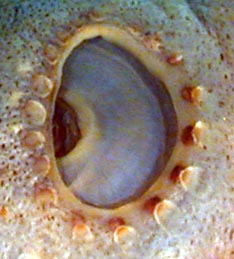
Figure. Left - Lateral views of left and right eyelids of H. atlantica, 53 mm ML. Drawings modified from Voss, 1969 (Fig. 32b, c). Right - Lateral view of right eyelid, 57 mm ML, 48°S, 145°E, USNM cat. no. 814656. Photograph by R. Young.
Click on an image to view larger version & data in a new window
Figure. Aboral view of arm I tip of H. atlantica, 34 mm ML male, 51° 59'S, 98° 59'E. Drawing from Voss, 1969 (Fig. 32h).
Click on an image to view larger version & data in a new window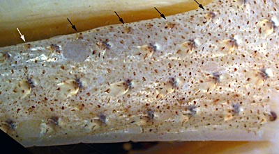
Figure. Ventral view of arm IV of H. atlantica, USNM no. 814656. Photograph by R. Young. The fourth series of small photophores is indicated by the arrows. At the arm base, smaller photophores can just be seen between the first four arrows.
Click on an image to view larger version & data in a new window
Figure. Ventral view of arm IV of H. atlantica, 53 mm ML. Extracted from Fig. 31a Voss, 1969.
- Tentacles
- Suckers on manus in about 6 series; 4 suckers in median series greatly enlarged with inner rings irregularly toothed (see drawing below).
Click on an image to view larger version & data in a new window
Figure. Oral view of club of H. atlantica, 53 mm ML. Drawing from Voss, 1969 (Fig. 31b).
- Arms
- Length 105-176% of ML (at 51-91 mm ML).
- Length 105-176% of ML (at 51-91 mm ML).
- Sucker dentition A- Large median club sucker; B- Sucker from row 2 arm II; C- Sucker from row 7 arm II. Sucker ring on bottom from large carpal sucker.
- Largest sucker rings from manal suckers irregularly incised, often partly smooth; dentition more regular in juveniles.
- Sucker rings of arms I-III vary from almost smooth to 5-10 low teeth on distal margin and occasionally on entire margin.
- Sucker rings of arms IV with more numerous, small, square teeth on distal or entire margin.
- Large carpal sucker may show accessory growth structure (drawing to the right, below).
Click on an image to view larger version & data in a new window
Figure. Oral view of sucker rings of H. atlantica, 53 mm ML, except carpal sucker which is from a 51 mm ML male, 35° 42'S, 18° 37'E. A - Largest tentacular sucker. B - Sucker from arm II row 2. C - Sucker from arm II row 7. Right - Ring of large carpal sucker at base of manus showing accessory growth. Drawings from Voss, 1969 (Fig. 32d-g).
- Web and buccal crown
- Inner web 17-30% of longest arm.
- Buccal crown with 7 supports.
Click on an image to view larger version & data in a new window
Figure. Oral view of buccal crown of H. atlantica, 53 mm ML. Drawing from Voss, 1969 (Fig. 32i).
- Funnel organ
- Dorsal pad with median ridge on each lateral arm
Click on an image to view larger version & data in a new window
Figure. Funnel organ, 34 mm ML of H. atlantica, 34 mm ML; Drawing from Voss, 1969 (Fig. 32j).
- Fins
- Length 26-47% of ML, width 53-65% of ML (at 51-91 mm ML).
- Length 26-47% of ML, width 53-65% of ML (at 51-91 mm ML).
- Spermatophore
- Short, ca 4-5% of ML; short sperm mass, 5-8% of spermatophore length (SpL); Cement body long, 75-80% of SpL; ejaculatory apparatus 14-17% of SpL with one major loop. Insert shows connection between ejaculatory apparatus and cement body. Note the unusual shape with a relatively short, slender oral end.
Click on an image to view larger version & data in a new window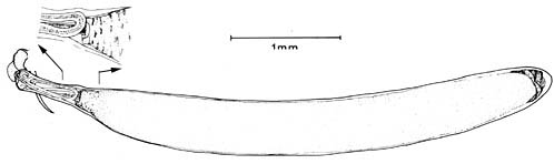
Figure. Spermatophore of H. atlantica, 87 mm ML, S. W. Atlantic. Drawing from Voss, et al., 1998.
- Short, ca 4-5% of ML; short sperm mass, 5-8% of spermatophore length (SpL); Cement body long, 75-80% of SpL; ejaculatory apparatus 14-17% of SpL with one major loop. Insert shows connection between ejaculatory apparatus and cement body. Note the unusual shape with a relatively short, slender oral end.
- Hectocotylus
- Apparently absent. Mature males with swollen, fleshy collars on basal suckers of all arms.
Comments
Except for the photographs, the above information is from Voss (1969) and Voss, et al. (1998).


